
Dooley Noted: 2/20/2015
Yesterday, I helped reflect 8 soleus muscles.
This thick, gorgeous leg muscle gets its name from its solefish-like appearance.
Failing to cross the knee, it joins the knee-crossing gastrocnemius at the tendocalcaneus (Achilles tendon) to insert into the heel.


Students are rightly nervous when reflecting this muscle.
They know that precious anatomy lays deep to its surface.
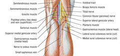
Soleus attaches to both leg bones, just inferior to the knee joint. It serves as a powerful plantarflexor of the foot, relative to the leg. Since it inserts medially into the heel, it also supports supination in the frontal plane.
In my opinion, soleus is often ignored when people have numbness of the foot. This is a mistake, since soleus can easily impinge the tibial nerve that supplies the sole of the foot.
Remember: soleus can impinge the sole’s sensation.
Soleus has an all-natural hiatus, allowing the passage of the tibial nerve and the posterior tibial artery. These crucial structures supply the posterior calf and the sole of the foot.
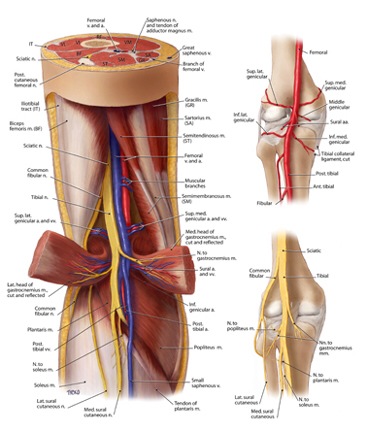
It’s at this hiatus that I become incredibly nervous while dissecting. There is a minute space available.
I say a quick prayer as I carefully push soleus between my blade and the neurovascular bundle passing tightly under the muscle.
As I carefully preserve the crucial neurovascular structures, I am amazed we don’t experience foot numbness and muscle cramping more than we do!
Soleus is mainly a one-joint muscle, leaving it to act largely on the ankle joint while supporting the subtalar joint. It’s also very close to the ground to provide stability when you lose it.
Thus, soleus tends toward facilitation, leaving you with a loss of dorsiflexion. This compensatory mechanism works to stabilize when structures higher up (I.e., core, hips) aren’t doing the job.
Like any compensation, the structure eventually fails.
Soleus overuse is functionally linked to everything from calf tightness to leg cramps to restless leg syndrome.
Even some cases of claudication (vascular occlusion) can be improved for comfort by addressing the soleus in the kinetic chain.
When stretching the soleus, the knee can remain bent since it doesn’t cross the knee.
But don’t be a newbie, going around stretching things that are attempting to stabilize you.
Consider releasing soleus of its increased tasks by finding ways to provide adjacent stability.
Get assessed for movement. Get corrected from soleus overuse or a lack of extensibility of this important muscle.
As always, it’s your call.
– Dr. Kathy Dooley